
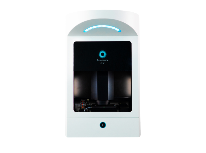
Tomocube HT-X1
-
Information
By capturing the intrinsic refractive index (RI) of cells using a low level of light intensity, Holotomography has emerged as a unique solution for live cell imaging that surpasses the compromise between obtaining high image quality and maintaining healthy cells.
The latest generation of Holotomography – Tomocube HT-X1 – utilizes a low-coherence light source. This groundbreaking advancement unlocks label-free 3D and 4D quantitative live cell imaging on diverse multi-well plates with improved resolution and free of laser-induced speckle noise.
The new system works well with a wide range of biological samples, including confluent cell sheets and thick specimens. It provides not only high-content 3D visualization but also quantitative data for individual cells and their intracellular structures, such as the volume, surface area, and dry mass.
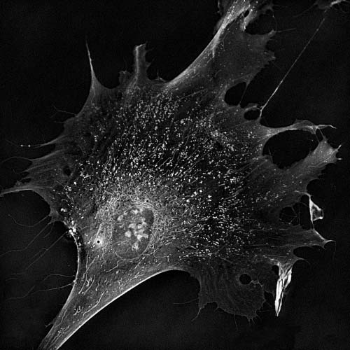
 3D visualization of monolayer cell cultures and 3D organoids
- Cellular and subcellular quantitative analysis
- Holotomography for label-free live cell imaging
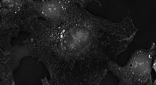
- Correlative fluorescence for biomolecular specificity
- Built-in incubator for long-term timelapse
- Multi-well plates compatibility for high-throughput experiment
- Fast, streamlined workflow for research efficiency
Tomocube HT-X1 utilizes a low-coherence LED light source to achieve speckle-free, high-contrast imaging without complicated alignments or calibration steps. This enables 3D imaging of thick samples, such as organoids.
A Digital Micro-mirror Device (DMD) is programmed to optimize the illumination patterns to encode specific spatial features of the specimen, providing high-content, high-resolution 3D imaging at exceptional stability.
Holotomography visualizes biological samples label-free based on their intrinsic RI properties, eliminating the need for long exposure to light. This minimizes phototoxicity and optimizes capture speed for 3D imaging to only 6.5 seconds per frame.
Moreover, an integrated stage-stop incubator allows stable environmental control for long-term timelapse observation and monitoring of cellular dynamics.
Especially designed to maximize user flexibility, the HT-X1 employs a unique adaptive illumination module that is tailored for multi-well plates. The combination of high NA, long working distance condenser, DMD, and a motorized illumination unit delivers an efficient illumination pattern for a diverse range of vessel types, from 35-mm dish to 96-well plate and even microfluidic chip.
With the HT-X1, researchers will have more freedom to design their experiments on different sample types using any imaging vessel of their choice.
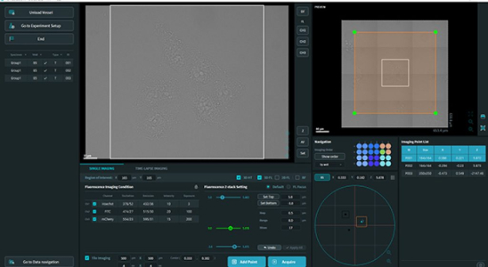
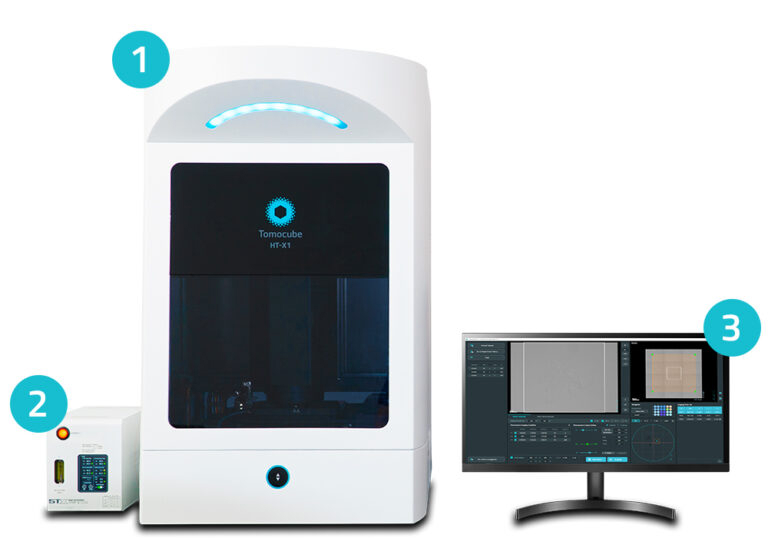
TomoStudioX works in concert with the HT-X1 platform to visualize and analyze RI tomograms. This intuitive, easy-to-use software empowers users with complete system control, allowing them to handle even the most complex experiments through simple mouse clicks.
- – Holotomography illumination
- – Detection
- – Sample holder
- – Environmental chamber
- – Positioning stages
- – Fluorescence light engine
- – External controller for temperature and gas
- – TomoStudio X for acquisition control
- – TomoAnalysis for data viewer & analysis